Evidence & Publications
At Cairina, our commitment to precision oncology is backed by cutting-edge research and validated methodologies. Our proprietary localized convolutional function regression (LCFR) technology transforms dynamic contrast-enhanced MRI (DCE-MRI) data into detailed, non-invasive maps of interstitial fluid dynamics, offering unparalleled insights into tumor physiology.

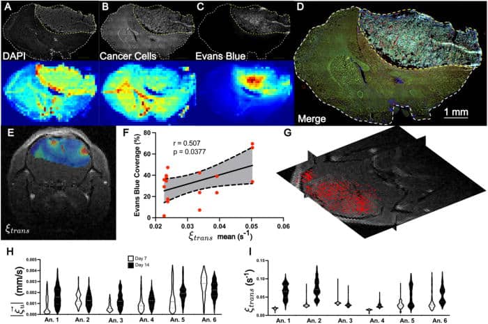
LCFR-measured perfusion is correlated with Evans Blue coverage in vivo. (a)–(d) Representative coronal IHC stains through the central tumor slice (top row) and MR resolution-matching intensity projection (bottom row), consisting of DAPI (a), GFP-expressing GL261 cells (b), Evans Blue (c). (d) Merge of all IHC demonstrating tumor heterogeneity. (e) Estimated perfusion field, ξtrans, overlaid on post-contrast T1-weighted image. (f) Scatter plot depicting the correlation and 95% confidence interval of linear regression between mean tumor perfusion as measured by DCE-MRI (ξtrans) and Evans Blue Coverage (mean tumor stain intensity), for N= 17 histology slices. (g) 3D velocity vector field of estimated interstitial velocity ξu→, overlaid on the 3D T1 post-contrast volume. (h) and (i) Violin plots depicting the estimated velocity magnitude (h) and perfusion (i) for six animals imaged 7 and 14 days after tumor implantation. (Read Study)

